A retinal detachment is a serious eye condition. The light-sensing tissue located at the back of your eye becomes detached from its supporting tissue, often due to a severe blow to the head. If not treated immediately, it can result in vision loss. Scleral buckling, though, can rescue the eye before eyesight deteriorates.
At Retina Specialists, our expert team of retinal ophthalmologists specializes in retinal conditions, and they offer scleral buckling among their treatment options for retinal tears and detachments. If you’re scheduled to go through this surgical procedure, here’s a step-by-step guide to what you need to know about it.
What is a retinal detachment?
Retinal detachment is an emergency situation where the retinal tissue pulls away from its moorings. That separates it from the blood vessels that provide it with oxygen and other nutrients. The longer you wait before getting medical help, the greater your risk of becoming permanently blind in the affected eye.
The detachment itself produces no pain, but it provides warning signs either before it occurs, or if it’s advanced, while it’s happening::
- Sudden appearance of visual floaters (dark specks in your vision)
- Bright flashes of light in one or both eyes
- Blurred vision
- Gradually reduced peripheral vision
- A curtain like shadow over your field of vision
If you experience any of these, call Retina Specialists to make an emergency appointment.
What is scleral buckling?
The only way to treat a retinal tear or detachment is surgically. A scleral buckling procedure repairs tears in the tissue and presses the retina against the wall.
A scleral buckle is composed of silicone sponge, rubber, or a semi-hard plastic. Your ophthalmologist places the buckle on the sclera, the white part of the eye, and sews it to the tissue to hold it in place. The buckle is generally placed permanently.
The material pushes the sclera, or "buckles it," toward the middle of the eye, relieving the pull on the retina and allowing the tear to rest against the wall of the eye. The doctor may choose to cover only the area of the eye behind the detachment, or they may choose to encircle the eyeball like a ring.
The scleral buckle by itself can’t prevent a retinal tear from opening again, so the doctor uses one of three methods to augment it: extreme cold (cryopexy), heat (diathermy), or light (laser photocoagulation). All of these scar the retina, holding it steady until a seal forms between the retinal tissue and the layer beneath it. The seal serves two purposes: 1) holds the layers of the eye together, and 2) prevents fluid from getting between them.
In most cases, a scleral buckle can reattach the retina and prevent further vision loss.
Your chances for good vision after the procedure are higher if the macula (the central 2% of the retina, which registers your clear, central vision) wasn’t part of the detachment. If the detachment did affect the macula, good vision is less likely.
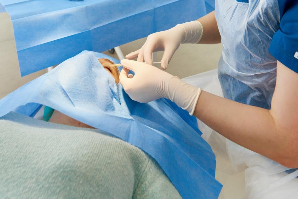
The scleral buckling procedure
It’s perfectly understandable to be anxious about your procedure; after all, it’s surgery. Fortunately, though, scleral buckling is highly successful. Here’s what you can expect during the procedure:
The doctor may give you anesthesia to put you to sleep. In this case, you won’t feel anything, and you won’t remember what happened afterward.
In some cases, you may remain awake, but the doctor gives you something to help you relax, and they’ll inject a local anesthetic, as well.
The surgeon puts dilating drops in your eye so they can better see all the structures, and then they expose the eye by making an incision in the outer layer, using an ophthalmoscope to get a clear view of the retina.
They seal the retina back together, most commonly using cryopexy. The retina is now attached to the inner wall of the eye.
Your surgeon places the buckle around the outside of your eye, much like you’d place a belt. This makes sure your retina stays in place.
They may drain some fluid from under the retina and probably will apply an antibiotic ointment to prevent infection.
Once the procedure is through and you’re alert, the team gives you a patch to cover up and protect your eye for a day or two.
If you’re experiencing any of the symptoms of a retinal detachment, make an emergency appointment with Retina Specialists to protect your vision. Call us at any of our Dallas, Texas, area locations.